Introduction
The research project Enhancement, classification and interpretation of histological images of cancer (Mejora, clasificación e interpretación de imágenes histológicas de cáncer) is part of the project Sistema de interpretación de imágenes histopatológicas para la detección de cáncer de próstata (SICAP) funded by the Spanish Ministerio de Economía, Industria y Competitividad from 2016 to 2020.
This is a coordinated project of the Computer Vision and Behaviour Anaysis Lab (CVLAB) from the Universidad Politécnica de Valencia and the Visual Information Processing Group (VIP) from the Universidad de Granada.
The Enhancement, classification and interpretation of histological images of cancer research team consists of 5 doctors from the University of Granada six undergraduates with extensive experience in the project theme and a doctor from the Northwestern University (Evanston, Illinois, USA).
This page will provide information on the project results and publications.
Summary
According to statistics, prostate cancer is the most common cancer among men in Spain. 32.641 new cases were diagnosed in 2014 (75% of those on patients over 65 years of age), being 30% the risk of having this type of cancer for men over 50. Most prostate cancers are first found during screening with a prostate-specific antigen (PSA) blood test or a digital rectal exam (DRE). However, the actual diagnosis of prostate cancer can only be made with a prostate biopsy.
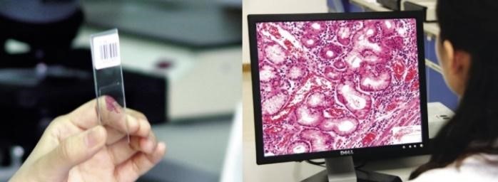
With the emergence of Whole-Slide Image (WSI) technology, high resolution scans of complete tissue biopsy slides are becoming a common clinical practice for prostate cancer diagnosis. However, in spite of the benefits of WSI for histopathological diagnosis, existing Computer Aided Diagnosis (CAD) systems primarily use only rectangular sections of WSI. Furthermore, commercial software tools for WSI analysis are currently very limited.
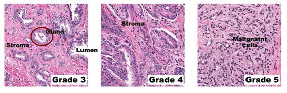
In this coordinated project, a multidisciplinary team of doctors, engineers, and scientists, some of them working at top foreign universities and research centers, will work together to develop new tools for prostate cancer diagnosis based on WSIs. Using publicly available databases and well as images acquired at the Hospital Clínico Universitario de Valencia, the team will work on image enhancement, automatic segmentation, tissue based feature extraction using doctors’ expertise and also on filter based feature extraction (no medical knowledge required). The extracted features will then be used on state of the arts classifiers for cancer diagnosis and grading using Gleason score. Together with the above classical approach to classification, Deep Learning (DL) techniques will, for the first time to the best of our knowledge, be used to perform the classification task. The initially best performing classification methods will mark the beginning of the loop: classification, expert’s interpretation, feature improvement and definition of new features, oriented to improve the system performance.
In summary, this project will research on and provide software for prostate cancer diagnosis and grading using WSIs. Results will be published at highly ranked journals and conferences. The software, whose commercialization will be sought, will improve WSI interpretation and help doctors in diagnosis tasks.
Objectives
The final objective of the project is the creation of a system that allows detecting the presence of cancer in a historical image providing, at the same time, an explanation of this decision in medical terms and identifying the regions and structures that support the decision.
O1. To obtain methods of improving the histological image that help to extract the most effective features and to increase the efficiency of the classification.
O2. To study and design a classification system based on features that determine the presence of cancer in an image and its degree of malignancy.
O3. To study and design a classification system without prior feature extraction (deep learning) that determines the presence of cancer in an image and its degree of malignancy as well as a strategy for a correct annotation in medical terms of the histological images classified as carcinogenic.